The presence of crystals in the urine is called crystalluria. Urinary crystals are usually detected on a urine test called “urinalysis.”
Having crystals in the urine is not always dangerous. Although these are very common and it’s normal to have a few tiny crystals in the urine. But in a few cases, they stick together and get big, called urinary stones. Sometimes it happens due to the storage of urine samples at room temperature because lower temperature favors stone precipitation.
The urine may have many components. It includes urinary crystals, RBCs, WBCs, squamous epithelial cells, transitional cells, neoplastic cells, urinary casts composed of mucoproteins mainly, and infectious agents like yeasts and bacteria.
What does it mean to have crystals in your urine?
Urine Crystals are usually described as highly organized microscopic solids that result due to the accumulation of different ions and/or molecules. Crystalluria, or having crystals in your urine, indicates the presence of too many minerals inside your urine.
Observing crystals in the urine is helpful in patients with known or suspected kidney stones and is a risk factor for recurrent calcium oxalate or cystine stone formation. In addition, crystalluria may have diagnostic utility in other settings:
- Magnesium ammonium phosphate crystals occur when there is an infection with urease-producing bacteria.
- The combination of calcium oxalate crystals and AKI may suggest ethylene glycol ingestion.
- The presence of AKI with uric acid crystals may suggest tumor lysis syndrome.
Factors causing Urine crystals
The presence of crystals in the urine depends upon the following factors
- Degree of concentration and solubility of crystallogenic substances in the specimen
- Excretion of diagnostic and therapeutic agents
- Evaporation, as it increases the solute concentration
- Urine pH as it changes with standing and bacterial overgrowth
- Temperature, as solubility decreases with temperature
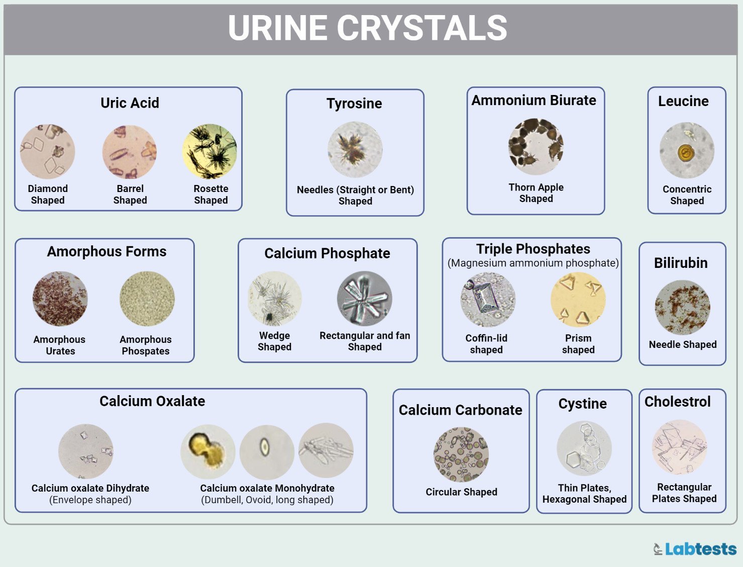
Types of urinary crystals
The urinary crystals have the following types.
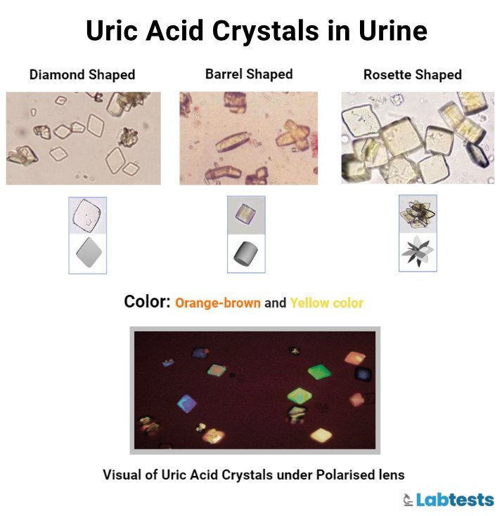
1. Uric acid crystals
- Uric acid crystals form in acidic urine.
- Microscope shows a pleomorphic appearance.
- Mostly they are like American football which means having slightly elongated shapes with rounded edges.
- Uric acid crystals are observed when there is hyperuricemia.
- The high uric acid content in blood and stones leads to crystal formation.
- These are usually of yellow-brown color.
- Commonly in leukemic patients on chemotherapy.
- Also seen in gouty arthritis, and tumor lysis syndrome.
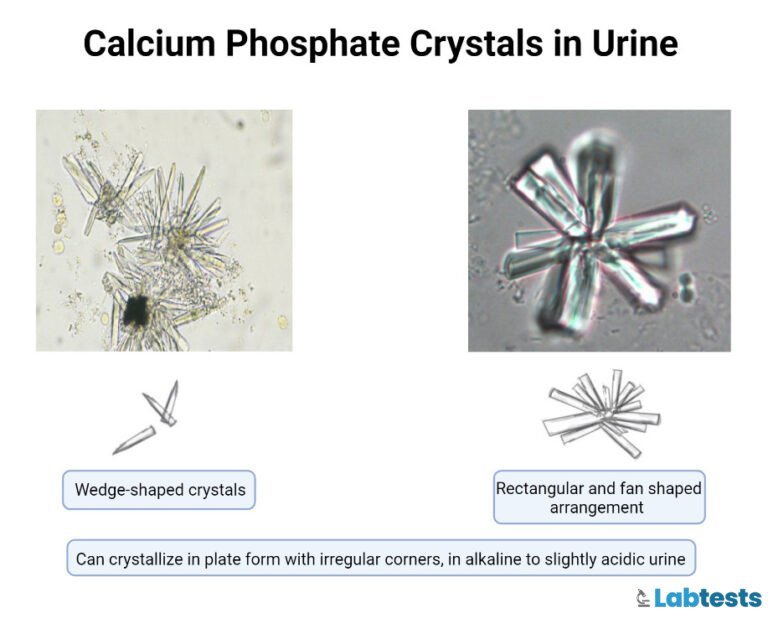
2. Calcium phosphate crystals
- These crystals form when the pH of urine is alkaline.
- It has a variety of shapes, including prisms that may merge into a rosette-like appearance.
- These are colorless.
- These are soluble in dilute acetic acid.
- These are involved in the formation of renal stones when present in huge quantities.
- If appeared on microscopy, these are less suggestive of any systemic disorder.
- These precipitate when pH increases and it’s due to the multiplication of bacteria.
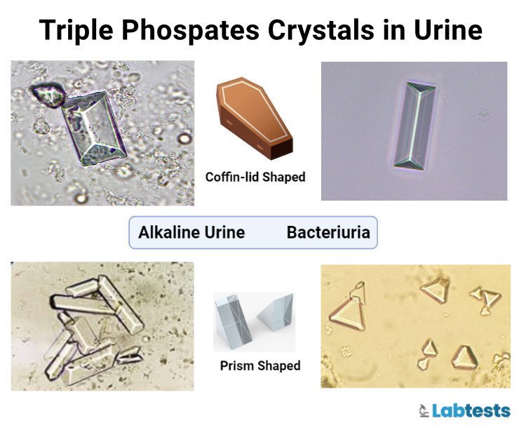
3. Magnesium ammonium phosphate crystals
- These are also called a struvite or triple phosphate crystals.
- Usually colorless, 3-dimensional, prism-like crystals (“coffin lids”) and occasionally resemble a double-edged razor blade.
- These may be present in the urine sample of clinically normal individuals.
- Urinary tract infections with organisms like urease-positive bacteria, such as Proteus or Klebsiella, can promote struvite crystalluria due to a rise in urine pH and increasing free ammonia.
- These can be present in the urinary sample of any pH but mostly in neutral to alkaline urine.
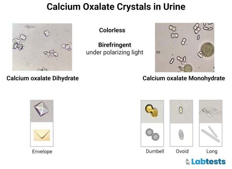
4. Calcium oxalate crystals
- These are colorless squares; their corners are linked by intersecting lines like an envelope.
- These do not depend upon the urine pH. More common in acid urine.
- Commonly when you take tomato, asparagus, ascorbic acid, etc.
- These vary in size and may appear in two forms
- 1) Monohydrate form rods, with a characteristic “dumbbell” appearance. Pleomorphic. These are also associated with ethylene glycol ingestion.
- 2) Dihydrate forms an envelope-like structure. It’s colorless. Rhomboids.
5. Cystine crystals
- These are flat, colorless plates with characteristic hexagonal shapes with equal or unequal sides.
- Their formation is favored in acidic urine and often aggregates in layers.
- These are seen in metabolic errors.
- In 1-2% of cases.
- These are soluble in ammonia and dilute HCL.

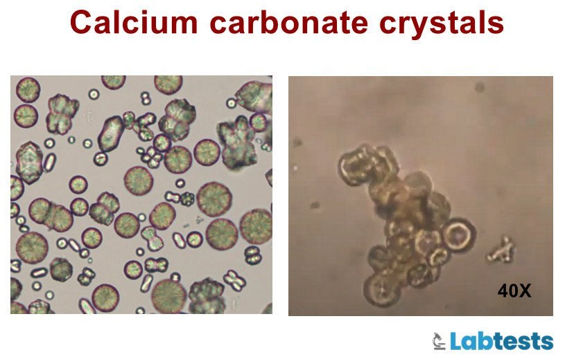
7. Calcium Carbonate crystals
- These usually appear in two forms.
- As large yellowish-brown or colorless spheroids having radial striations.
- As smaller crystals with oval, round, or dumbbell shapes.
- If acetic acid is added, there will be gas formation.
- These are seen in an alkaline medium.
- Calcium carbonate crystals are huge circular discs with smooth surfaces characterized by colorless to light brown color. Although they may also appear smaller in size.
- A high concentration of calcium carbonate crystals in the urine causes the urine to be brownish-tinged.
- Calcium carbonate crystals are commonly seen in the urine of rabbits, goats, horses, and guinea pigs.
- These crystals shouldn’t be seen in human urine as well as in the urine of cats and dogs.
- The presence of calcium carbonate crystals in urine is usually caused by excessive intake of calcium supplements such as crystals of calcium carbonate.
- If your urine contains calcium carbonate crystals, your doctor will advise you to discontinue calcium supplements and get your daily need for calcium from other sources such as through your diet.
8. Bilirubin crystals
- Bilirubin is a product of the healthy destruction of red blood cells.
- These fine needle-like crystals precipitate onto other elements in the urine.
- These crystals look like needles, look like tiny granules, and are yellow in color.
- The other less common shape is cylindrical, which is formed in association with droplets of fat, resulting in a “flashlight” appearance.
- The presence of bilirubin in urine indicates poor liver function and possible liver-related disease, especially when accompanied by other alarming symptoms like nausea, vomiting, fever, pain, and jaundice.
- The treatment management depends on the underlying cause but usually includes medication and lifestyle changes.
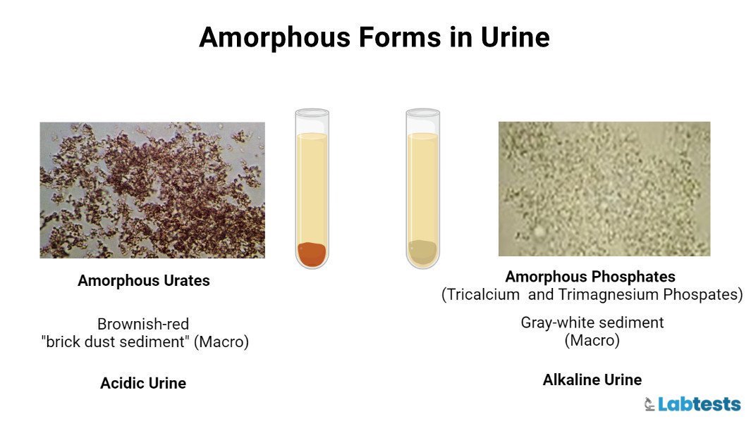
9. Amorphous Forms
- These are aggregates of finely granular material and don’t have any defined shape at the microscopic level.
- Urates (Mg, Na, K, or Ca salts) form in acidic urine and have a yellow or yellowish-brown color.
- In general appearance, phosphates are similar but alkaline urine favors formation. These are colorless. Granular in shape.
- Calcium oxalate dihydrate crystals can sometimes be “amorphous” when the individual crystals are tiny. Examination at higher magnification will reveal the typical “envelope” appearance.
10. Drug crystals
Some drugs excreted in the urine have the potential to form crystals.
The most common among these drugs are sulfa drugs, acyclovir, sulfonamides, atazanavir, and methotrexate.
| Drug | Crystal | Clinical manifestations |
|---|---|---|
| Sulphadiazine | Birefringent “shocks” of wheat or “shells” with striations (Image) | Isolated crystalluria, haematuria, ARF, Stones |
| Acyclovir | Birefringent fine needles (Image) | Isolated crystalluria? ARF |
| Indinavir | Plate-like rectangles, star-like forms, irregular plates, strongly birefringent (Image) | Isolated crystalluria, stones, ARF |
| Piridoxylate | Asymmetrical hexagons of rectangles with rounded extremities | Stones |
| Primidone | Birefringent hexagons | Isolated crystalluria, transient proteinuria and haematuria |
| Naftidrofuryl Oxalate | Birefringent monohydrate calcium oxalate (Image) | ARF |
| Vitamin C | Birefringent monohydrate calcium oxalate (Image) | Acute renal failure |
| Amoxycillin | Needles Shocks of wheat (Image) | Isolated crystalluria Haematuria ARF |
Research Laboratory on Urine, Unità Operativa di Nefrologia
Fondazione IRCCS, Ospedale Maggiore Policlinico, Mangiagalli e Regina Elena
Milan, Italy)
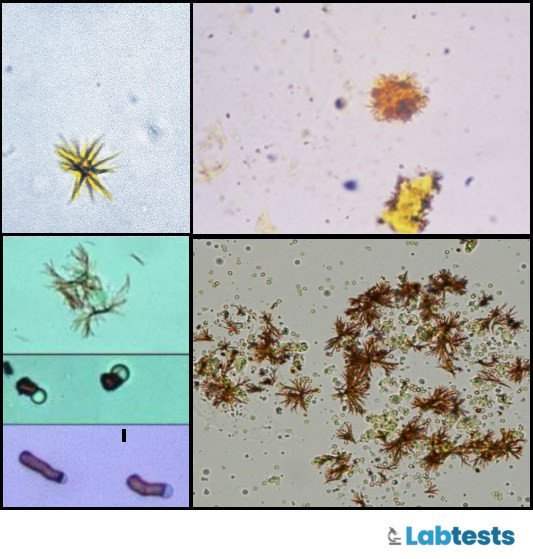
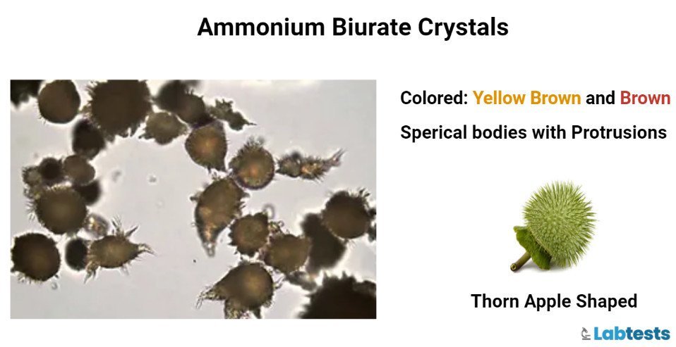
11. Biurates (Ammonium urate) crystals
- Ammonium urate, also called biurate crystals, generally appear as brown or yellow-brown spherical bodies with irregular protrusions (“thornapples”).
- These are possible in the urine of any pH, but the formation is favored in neutral to alkaline urine.
- These dissolve at 60 ℃.
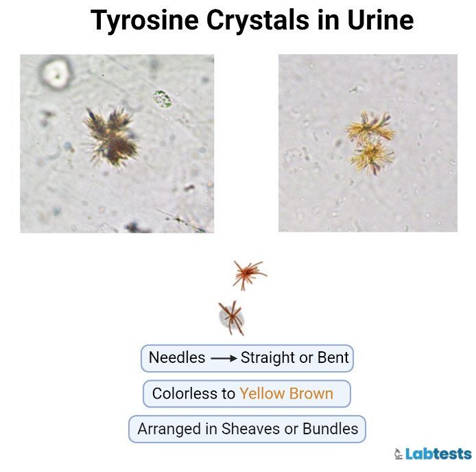
12. Tyrosine crystals
- These are usually seen as fine brownish needles and can be associated with severe liver disease.
- These are fine colorless yellow needle-shaped crystals.
- Seen in inherited disorders of amino acid metabolism.
- These are soluble in alkali or by heat.
13. Leucine crystals
- Leucine crystals also known as wagon wheels are found in acidic urine and characterized by yellow-brown color and concentric rings that resemble a tree trunk.
- Leucine crystals in the urine are a result of detection in leucine (amino acid).
- They form in neutral/acid medium.
- These crystals are soluble in alkali and hot alcohol.
- Leucine crystals are accompanied by tyrosine crystals but are less frequently seen than the latter.
- Leucine crystals in urine indicate severe liver disease, especially when accompanied by other alarming symptoms like nausea and vomiting, general body malaise, disorientation, and abdominal swelling.
- The focus of treatment is to improve liver function and overall health. the patient receives all pertinent medication with the goal of reducing swelling and reducing the possibility of bleeding.

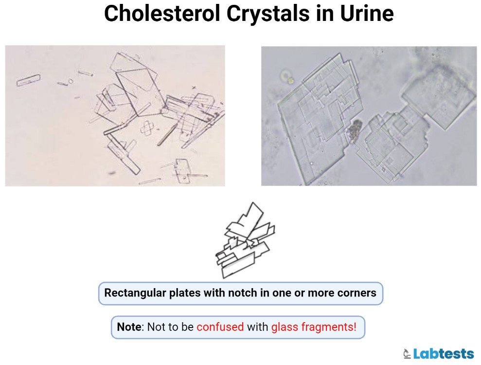
14. Cholesterol crystals
- For cholesterol crystals to be detected in urine, the urine must be refrigerated first.
- Cholesterol crystals look like rectangular plates with a noticeable notch (cut-out) in the corners.
- They appear in acidic medium, soluble in chloroform and ethanol medium but insoluble in alkalis and acids
- These crystals can appear in both acidic and neutral urine.
- They can only be found in the urine of people with severe kidney disease, ruptured lymphatic vessels in the renal pelvis, and renal tubular disease leading to renal failure.
- Treatment includes alkali therapy to address problems like renal tubular disease and other related chronic metabolic conditions.
List of Urine Crystals and Shapes (Pictures)
Crystals in the acidic urine
| Name of crystals | Color | Shape | pH | Effect of heating (solubility) |
|---|---|---|---|---|
| Ampicillin | Colorless | in the form of needles | Acidic pH/ neutral pH | – |
| Calcium oxalate | Colorless octahedral envelope/ two pyramids joined at the base. | enveloped shaped, dumbbell-shaped | Acidic pH or alkaline pH | dilute HCL |
| Uric acid | yellow-brown | rhomboid, four-sided flat plates | Acidic pH <5.5 | Alkali- soluble |
| Amorphous urates | Microscopically yellow-brown and occurs in clumps | amorphous or sand-like | acidic pH >5.5 | alkalies and heat |
| Sodium urates | Acidic pH | |||
| Sulphonamide | colorless to yellow-brown | Rosette form, needle, | Acid pH/ neutral | Acetone |
| Cholesterol | Colorless | rectangular, notched plates | Acidic pH | Chloroform |
| Cystine | Colorless | hexagonal | Acidic pH | Dilute HCl acid |
| Amorphous urates | Microscopically yellow-brown and occurs in clumps | amorphous or sand-like | Acidic pH >5.5 | alkalies and heat |
| Tyrosine | Colorless to yellow, needles | needles shape form clumps or rosettes | Acidic pH / neutral | Alkali or heat |
| Bilirubin | yellowish | clumped needles or granular | Acidic pH | Acetic acid, HCL,ether or chloroform |
| Radiographic dye | colorless | like cholesterol | Acidic pH | 10% NaOH |
| Sodium urate | Colorless to yellow, Amorphous, or having large granules | amorphous | Acidic pH | soluble at 60 C |
| Leucine | Yellow-brown Color | spheres with a concentric circle or radial striations | acidic pH/ neutral | Hot alkali or alcohol |
Crystals in alkaline urine
| Name of crystals | Color and shape | Effect of heat | pH |
|---|---|---|---|
| Calcium Carbonate | Small, colorless with a dumbbell or spherical shape, may occur in clumps and resemble amorphous material | Alkaline | |
| Triple phosphates | Colorless and prism-shaped like a coffin lid | Alkaline | |
| Amorphous phosphates | Granular | Remains insoluble | Alkaline |
| Calcium phosphate | Colorless, flat rectangular plates or thin prisms often in rosettes | Remains insoluble | Alkaline |
| Ammonium biurate | Yellow-brown colors, thorny apple | Dissolves at 60 °C | Alkaline |
The Solubility Of Crystals
| Crystals | pH | Color | Soluble in |
|---|---|---|---|
| Calcium oxalate | Acidic /neutral | Colorless | Dilute HCl acid |
| Amorphous urates | Acidic | Brick dust or yellow-brown | Alkali and when heated |
| Ammonium biurate | Alkaline | Yellow-brown | Acetic acid with heat |
| Triple phosphate | Alkaline | Colorless | Dilute acidic acid |
| Uric acid | Acidic | Yellow-brown | Alkali |
| Calcium carbonate | Alkaline | Colorless | Acetic acid |
| Calcium phosphate | Alkaline/neutral | Colorless | Dilute acidic acid |
| Amorphous phosphate | Alkaline/neutral | White-colorless | Dilute acid acid |
Causes of urinary crystals
Some of the most common causes of urinary crystals are the following
- Dehydration
- Some medicines like amoxicillin, acyclovir, methotrexate, and sulfonamide
- UTI
- Ethylene glycol poisoning
- Tumor lysis syndrome
What is the clinical significance of Urinary crystals?
Urinary crystals are clinically significant and associated with many medical conditions.
- Calcium oxalate crystals are associated with nephrolithiasis, ethylene glycol ingestion, and excessive oxalate absorption.
- Calcium phosphate crystals are associated with nephrolithiasis in patients with defects of urine acidification.
- Triple phosphate or struvite crystals are associated with infection with urea-splitting organisms.
- Uric acid crystals are associated with nephrolithiasis and tumor lysis syndrome.
- Cystine crystals are associated with cystinuria.
- Urinary crystals are also crucial in renal damage, liver diseases, and metabolic errors.
- Transient supersaturation is one of the common reasons for crystalluria.
- Crystalluria can also be due to the ingestion of certain foods.
Precautions for urinary crystals
- Past history of drug/medication use is essential.
- pH is also important to be noted.
- Refrigeration affects and precipitates many crystals. It changes the solubility.
- Temperature also affects (i.e. samples kept at room temperature dissolves crystals).
- Radiographic dye is involved in crystal formation especially when the patient is dehydrated.
- Some medications used like ampicillin and sulfonamides are involved in crystal formation, especially in dehydrated patients.
Frequently asked questions
Q1: What are the signs and symptoms of urinary crystals?
The common signs and symptoms are
• Pain
• Fever
• Blood in urine
• Urinary obstruction
• Urinary frequency
Q2. How can I avoid urinary crystal formation?
These simple techniques can lower the risk of urinary crystals.
• Avoid taking medications without a prescription.
• Drink an adequate amount of fluid
• Maintain healthy weight
• Take a moderate amount of proteins
• Take an average daily intake of vitamin C, calcium, and oxalate.
• Avoid taking too much salt and sugar.
Q3. What is the specimen of choice for urinary crystals?
The specimen of choice to evaluate urinary crystals is
• First/second-morning, midstream, and clean-catch urine sample. Although the first-morning urine is the most concentrated for microscopically examining cells, the second-morning specimen is preferred. Because in the first-morning sample, the cellular elements can be damaged or decomposed.
• The urine volume should be 6-12 mL.
• The sample should be fresh and taken within one hour.
Q4. What is the role of refrigeration in urinary crystals?
The refrigeration process will precipitate the formation of the crystals. It is due to the change in solubility of various crystals.
References
- Vijaya, T., Kumar, M. S., Ramarao, N. V., Babu, A. N., & Ramarao, N. (2013). Urolithiasis and its causes-short review. J Psychopharmacol, 2(3), 1-6.
- Worcester, E. M. (1994). Urinary calcium oxalate crystal growth inhibitors. Journal of the American Society of Nephrology, 5(5), S46.
- Crystals in Urine: Types, Causes, Symptoms & Treatment. (n.d.). Cleveland Clinic. Retrieved October 16, 2022, from
https://my.clevelandclinic.org/health/diseases/22204-crystals-in-urine - Gotter, A. (2020, June 10). Crystals in the Urine: What You Need to Know. Healthline. Retrieved October 16, 2022, from https://www.healthline.com/health/urine-crystals